
Cone Beam CT for Rodents – Modern Diagnostics
Dr. med. vet. Cornelia Christen, Dipl. ECZM
31.08.2025
Cone Beam CT for Rodents – Modern Diagnostics for Sensitive Patients
By Dr. med. vet. Cornelia Christen, Dipl. ECZM
Small pets such as rabbits, guinea pigs or chinchillas are masters at hiding pain. Dental problems in particular often go unnoticed for a long time because the animals do not show obvious signs of discomfort. To detect health changes at an early stage, diagnostics are needed that reveal even the smallest details while taking into account the animals’ special needs. Cone Beam Computed Tomography meets exactly these requirements.
What is Cone Beam CT and what is its advantage for rodents?
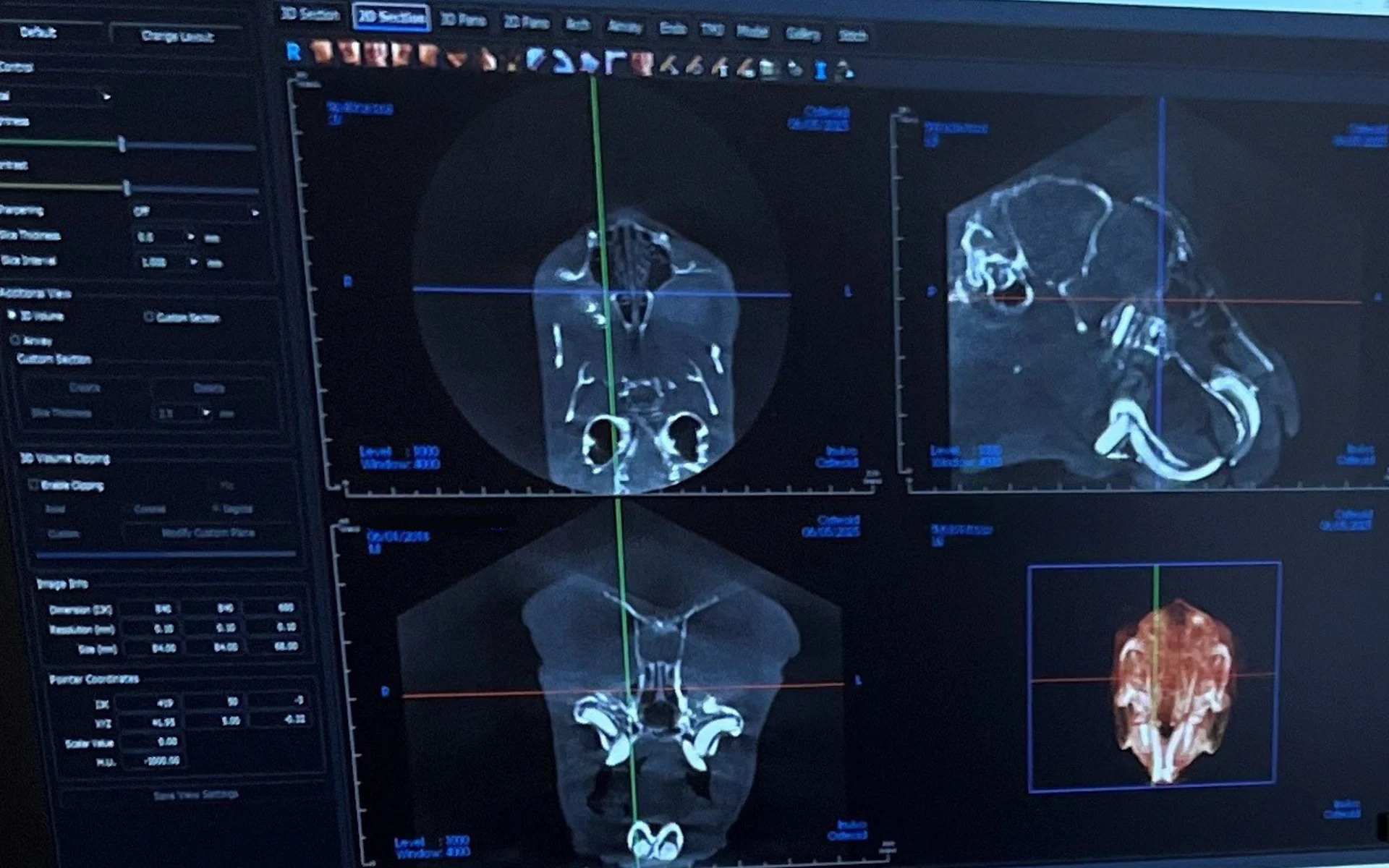
Cone Beam CT is a modern imaging technique that enables three-dimensional, high-resolution images. The key difference from conventional computed tomography lies in the technology: instead of scanning the body layer by layer with a fan-shaped beam, Cone Beam CT uses a cone-shaped X-ray beam that captures the desired area completely in a single rotation. This allows precise images to be produced with a significantly lower radiation dose.
For rodents, whose anatomical structures are particularly fine and closely spaced, this method offers crucial advantages. It provides clear images of the skull, teeth, jaw, and nasal cavities – with minimal stress for the animal.
When can Cone Beam CT be useful for small rodents?
Many head diseases in small pets are hardly recognisable from the outside. Initial signs such as refusal to eat, drooling or weight loss are often mistaken for general discomfort. In fact, dental diseases or inflammations are often behind these symptoms and can only be reliably detected using high-resolution imaging.
Cone Beam CT is particularly useful for:
- Changes in tooth roots
- Suspected jaw abscesses or cysts
- Chronic nasal problems
- Asymmetrical growth or jaw misalignment
- Unclear causes of recurring feeding problems
It is also a reliable basis for planning interventions or for follow-up after surgery.
Cone Beam CT or conventional CT?
Both methods have their merits but differ in application. Conventional CT provides good results for soft tissue imaging but takes more time and involves higher radiation exposure. For small pets where the focus is on teeth, bones, and fine head structures, Cone Beam CT is often the better choice.
Its advantages:
- Short examination time
- Lower radiation exposure
- High-resolution 3D images
- Less stress due to shorter anaesthesia
- Ideal for animals with delicate anatomical structures
Cone Beam Computed Tomography
Procedure and care: what will my guinea pig or rabbit experience at VetTrust?
Before any imaging, our team carries out a thorough examination. Together with the owners, we decide whether Cone Beam CT is necessary and appropriate. The scan itself is performed under short anaesthesia to keep the animal still and safe. Our specialists accompany the entire process – from preparation to the recovery phase.
After the examination, our experienced veterinarians evaluate the images and discuss the next steps in detail. The goal is always to provide treatment that helps the animal as quickly as possible while avoiding unnecessary procedures.
Our approach: technology is only as good as the person using it
At VetTrust, we use modern methods only when they provide real added value for our patients. Cone Beam CT is an example of how precise diagnostics, veterinary expertise, and compassion can go hand in hand. Our smallest patients deserve the same attention and care as the bigger ones.
If you are unsure whether a more detailed examination is appropriate or whether Cone Beam CT is the right choice, we will be happy to advise you.
Book your appointment now with Dr. med. vet. Cornelia Christen at VetTrust Small Animal Practice Winterthur.