Cruciate ligament tear in dogs
Dr. med. vet. Karol Bayer, Dipl. ECVS
31.08.2025
Cruciate ligament tear in dogs: causes, symptoms and modern treatment options
When dogs suddenly limp or stop putting weight on a hind leg, a cruciate ligament tear is a common cause. It is one of the most frequent orthopedic conditions in dogs and can affect any size and age. It helps owners to understand the causes, recognize typical signs and know the treatment options.
What happens in a cruciate ligament tear
Two important ligaments run through the dog’s knee joint, the cruciate ligaments. They stabilize the joint during movement. The front cruciate ligament is particularly prone to injury. If this ligament tears completely or partially, the femur can no longer glide correctly on the tibia. The knee becomes unstable and painful, and dogs adopt protective postures. Without treatment this can lead to joint wear and tear, also called osteoarthritis.
How a cruciate ligament tear develops
Unlike in people, where sports accidents are common, most cruciate ligament tears in dogs have a degenerative origin. The ligament tissue loses elasticity and normal function over time. Partially torn cruciate ligaments do not heal back together and the damage progresses.
Often several factors are involved:
- Wear of the ligament tissue over time
- Unfavorable knee joint angulation
- Excess weight
- Breed predisposition, for example Labrador Retriever or Rottweiler
- Misloading due to other orthopedic problems
- Less commonly, acute trauma while playing, running or jumping
Many affected dogs first show occasional lameness. Signs often increase over time, especially after rest or exercise.
How to recognize a cruciate ligament tear
A complete tear often causes sudden, marked lameness of the affected leg. With partial tears, symptoms develop more slowly and may be missed. Typical signs include:
- Lameness or inconsistent weight bearing of the hind leg
- Stiffness or limping after getting up that improves with movement
- Unsteady gait or visible knee instability
- Avoiding stairs or jumps
- Thickening or swelling of the knee joint
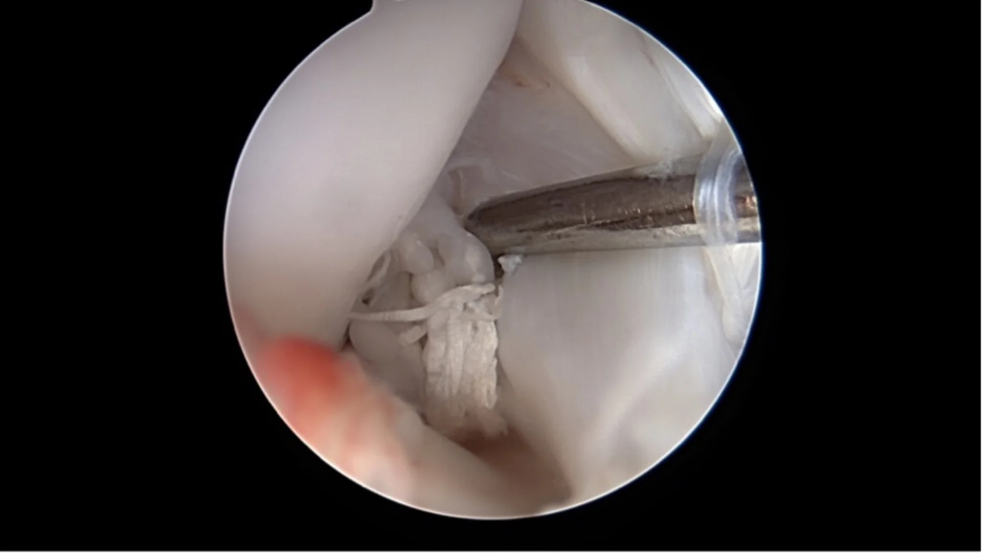
An incipient cruciate ligament tear is hidden beneath the remaining cruciate ligament, which appears unremarkable at first glance.

A complete cruciate ligament rupture
A veterinarian can confirm the diagnosis with targeted motion tests and imaging.
How is a cruciate ligament tear treated
Treatment depends on your dog’s age, weight, activity level and overall health. In almost all cases surgery is recommended to restore joint stability and prevent secondary damage. Early diagnosis and treatment reduce the risk of additional injuries such as meniscal tears or cartilage damage and support faster recovery.
Surgical options at a glance
TPLO, Tibial Plateau Leveling Osteotomy
The slope of the tibial plateau is surgically altered so that the load axis of the knee changes. This neutralizes the stabilizing role of the front cruciate ligament without replacing it. Based on the growing body of objective studies, TPLO is the only method shown to restore load bearing to the level of the healthy limb. It is considered the international gold standard in cruciate surgery for dogs and is suitable for all sizes.
The slope of the tibial plateau is surgically altered so that the load axis of the knee changes. This neutralizes the stabilizing role of the front cruciate ligament without replacing it. Based on the growing body of objective studies, TPLO is the only method shown to restore load bearing to the level of the healthy limb. It is considered the international gold standard in cruciate surgery for dogs and is suitable for all sizes.
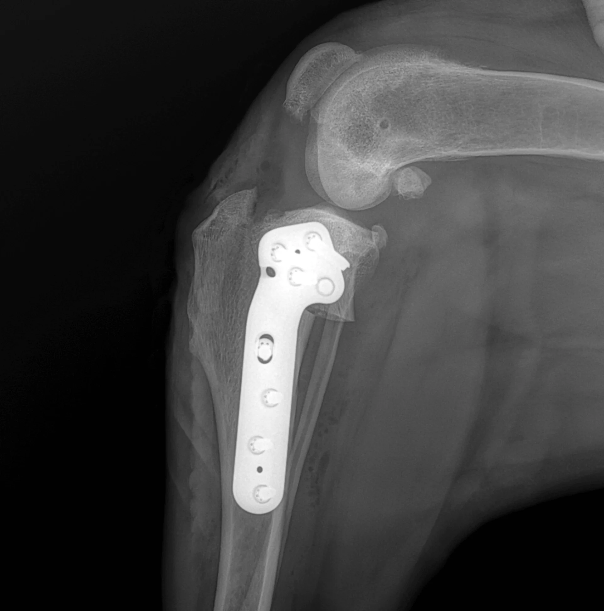
Postoperative X-ray image of the TPLO technique. The rotated tibial plateau, which is fixed with a special plate, can be seen.
TTA, Tibial Tuberosity Advancement
This procedure changes the direction of pull of the patellar tendon. The function of the cruciate ligament is replaced by higher tension in the patellar tendon. It can help selected patients. Recent studies showed variable outcomes, which calls the consistency of the intended mechanism into question. It is therefore no longer considered the global gold standard for cruciate surgery in dogs.
This procedure changes the direction of pull of the patellar tendon. The function of the cruciate ligament is replaced by higher tension in the patellar tendon. It can help selected patients. Recent studies showed variable outcomes, which calls the consistency of the intended mechanism into question. It is therefore no longer considered the global gold standard for cruciate surgery in dogs.
Extracapsular ligament replacement
A synthetic suture is placed outside the joint along the course of the front cruciate ligament to provide passive stability. Stability usually declines after several months while fibrous tissue forms that can help stabilize the joint. This method is mainly suitable for cats and very small patients.
A synthetic suture is placed outside the joint along the course of the front cruciate ligament to provide passive stability. Stability usually declines after several months while fibrous tissue forms that can help stabilize the joint. This method is mainly suitable for cats and very small patients.
Gentle and precise: minimally invasive techniques
Arthroscopy
A miniature camera allows direct visualization of the inside of the joint through small skin incisions. Changes to the cruciate ligament and associated damage such as meniscal tears can be diagnosed and in many cases treated immediately.
A miniature camera allows direct visualization of the inside of the joint through small skin incisions. Changes to the cruciate ligament and associated damage such as meniscal tears can be diagnosed and in many cases treated immediately.
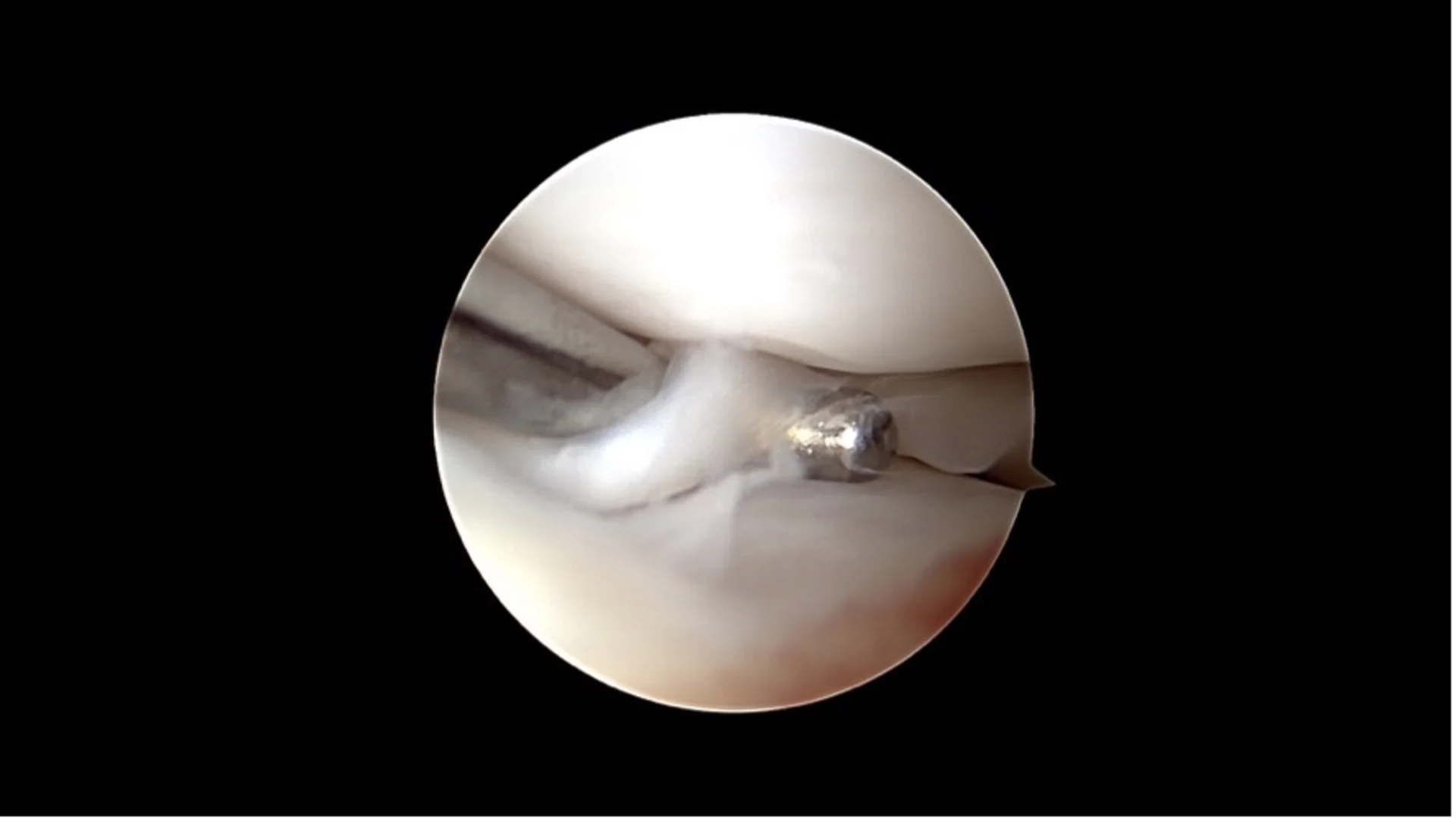
Basket-handle meniscus tear
Nanoscopy
Even finer than arthroscopy, nanoscopy uses very small instruments and high resolution imaging inside the joint. It is particularly suitable for small dogs. Both methods are minimally invasive, cause less tissue trauma and support faster recovery.
Even finer than arthroscopy, nanoscopy uses very small instruments and high resolution imaging inside the joint. It is particularly suitable for small dogs. Both methods are minimally invasive, cause less tissue trauma and support faster recovery.
What to expect after surgery
Aftercare is essential. Recovery depends on how consistently the rehabilitation plan is followed. Typical elements include:
- Controlled, calm movement for about 6 to 8 weeks
- Wound care with an Elizabethan collar until skin healing about 10 days after surgery
- Controlled weight bearing with leash support and bandages if needed
- Physiotherapy for advanced cases with muscle atrophy
- Pain management and anti inflammatory therapy
Depending on the procedure and the individual case, full weight bearing is usually expected after about eight to twelve weeks. A final checkup, possibly including X rays, is scheduled.
Good prognosis with timely treatment
A cruciate ligament tear is painful but usually very treatable. Early examination and expert care are key. With today’s surgical and minimally invasive techniques, long term outcomes are very good when diagnosis, therapy and aftercare work hand in hand.
If you suspect a cruciate ligament tear in your dog, a thorough examination provides clarity and options for targeted help so your dog can move safely and without pain again.